Sunday, December 8, 2013

My Dictionary: Urinary System

Cortical labyrinths- the region between medullary rays contains renal corpuscles

http://www.dartmouth.edu/~anatomy/Histo/lab_5/renal/DMS152/popup.html

Bowman's capsule- double layered epithelial cup 

http://www.ouhsc.edu/histology/text%20sections/urinary.html
 Juxtamedullary nephrons- 1/8 of total nephron count. Contain long loops of Henle that go into the inner region of the pyramid.

http://faculty.southwest.tn.edu/rburkett/A%26P2%20urinary_system.htm

Sunday, December 1, 2013

Reflections: Digestive System III

     The liver is surrounded by fibrous connective tissue that is called Glison's capusle. Exterior to the capsule is a serous covering, the visceral peritoneum. However, the visceral peritoneum is not found where the liver meets with the diaphragm and other organs. The liver has grooves that divided into four lobes, two large lobes (left and right lobe) and two smaller lobes (quadrate and caudate lobes). The liver starts off as an endodemal evagination in the embryo. Then it forms the hepatic diverticulum, which proliferates and create hepatocytes that constitute the parenchyma. The liver is important in producing and secreting plasma proteins, regulating vitamins and nutrients, and maintains blood glucose levels. Moreover, it also plays a role in degrading drugs and toxic substances. The liver acts as an exocrine gland because it produces bile secretion and lastly, it also functions as an endocrine gland.
     The gallbladder is located at the visceral surface of the liver. Its main function is to store bile, it also removes approximately 90% of the water. The small intestine secretes hormones that cause contractions of the gallbladder that lead to bile discharge into the duodenum. The pancreas is divided into three sections: head, body, and tail. The gland is surrounded by a capsule that is made up of a layer of connective tissue. The pancreas as both an endocrine and exocrine gland. The endocrine component of the gland regulates blood glucose levels by synthesizing and secreting insulin and glucagon. The exocrine component of the gland functions as a serous gland and secretes digestive enzymes. 

Wednesday, November 20, 2013

Salivary Gland Tumors

     Salivary gland tumors typically occur in the major salivary glands. The three major salivary glands are the parotid gland, submandibular gland, and sublingual gland. The minor glands are found throughout the mouth, pharynx, larynx, and paranasal sinuses. Most of the salivary tumors are benign, meaning that they are not cancerous. There are usually in the parotid gland or in the palate if it is a minor salivary gland tumor.
    Most of the benign tumors are pleomorphic adenoma tumors. This tumor has epithelial tissue with ducts and epithelial cells mixed with areas that have similar characteristics of ground substance of connective tissue. The myoepithelial cells are what produce the tissues that resemble connective tissue. Patients that have these benign tumors claim to have no pain in the affected glands. The reason for this may be that the innervated nerves are numb and weak. What should a patient do with a benign salivary gland tumor? Surgery is a possible option for parotid gland tumors in which the entire gland will be removed.
Pawlina, Wojciech. "Connective Tissue." Histology A Text and Atlas By Michael H. Ross. 6th ed. N..: n.p., n.d 555 Print.               The two main contributors listed for the 6th edition of this textbook, Wojciech Pawlina and Michael H. Ross.
http://emedicine.medscape.com/article/194522-overview

My Dictionary

Palatine tonsils- located on both sides of the oropharynx opening
http://www.webmd.com/oral-health/picture-of-the-tonsils
Tubal tonsils- located behind the auditory tube opening on the later walls
Lingual tonsil- located at the tongue's base

http://www.knowyourbody.net/tubal-tonsil.html

Pharyngeal tonsils- at the top of the nasopharynx
http://hanatomy.com/anatomy-directory/lingual-palatine-and-pharyngeal-tonsils/

Thursday, November 14, 2013

Reflections: Integumentary System


     As we closed the chapter dealing with the integumentary system, I decided it would be good to do a brief review on what we learned. The integumentary system is composed of two main layers: the epidermis and the dermis, and there are two types of skin (thin and thick). Thin skin has hair and is more prevalent as opposed to thick skin which is only found in the palms of the hands and the soles of the feet.
     The epidermis is the layer of skin that is exposed and is visible. The dermis lies directly beneath the epidermis. Then there is the hypodermis that contains adipose tissue and connective tissue. The epidermis is further divided into 5 layers: stratum corneum, stratum lucidum, stratum granulosum, stratum spinosum, and stratum basale. The stratum corneum is the layer that varies in thick skin.
     The cells of the epidermis are keratinocytes, melanocytes, Langerhans', and Merkel's cells. The epidermis is made up of keratinized stratified squamos epithelium. The keratinocytes are the cells that function to separate the organism from the outside environment. Melanocytes are the cells that secrete melanin. Melanin is important because it is the reason that we are protected from UV radiation. It also produces the pigment of the epidermis. Langerhans' cells are the one's that present antigen's to the immune system. Lastly, Merkel's cells function in stratum basale of the epidermis and they are the most abundant in the fingertips and other places that sensory perception is intense.
http://classroom.sdmesa.edu/eschmid/Chapter5-Zoo145.htm

Thursday, October 31, 2013

Encounters: Urinary Tract Infection

      
     After finishing the lymphatic system chapter and turning in our disease projects this week, I felt motivated to write about a situation that happened to my friend. Earlier this week, she was experiencing back pain and had a burning sensation during urination. She was soon diagnosed with a urinary tract infection. Since she is my friend, I was worried about her and felt compelled to research the disease itself, the causes, and the symptoms. 
     In my research I discovered that UTI's are basically bacteria that has infected the urinary system. What entirely does the urinary system comprise? It consists of the kidney's, ureters, bladder, urethra, and sphincter muscles. Since women have short urethra's they are more susceptible to getting a UTI than men. Causes vary greatly and range from having sexual intercourse to pieces of feces coming into contact with the urethra. It can happen whenever bacteria gains access to the urethra in any way or form. 
Symptoms may include the following:

  • cloudy urine
  • painful/ burning sensation when urinating
  • frequent need to urinate
  • back pain
  • abdominal pain
  • nausea
     Fortunately, there are antibiotics available that will help with the discomfort. However, after taking the antibiotics she did mention that she felt extremely nauseous, dizziness, and wanted to vomit. She is now well and not feeling any discomfort. 


http://www.medicalnewstoday.com/articles/189953.php
http://www.mayoclinic.com/health/urinary-tract-infection/DS00286/DSECTION=symptoms
http://www.plannedparenthood.org/health-topics/womens-health/utis-4283.htm

Tuesday, October 29, 2013

Human Immunodeficiency Virus

     HIV is a retrovirus and it attacks the immune system by invading T helper cells. It contains an enzyme called reverse transcriptase that aids it in making DNA with the RNA that it has. After several years, approximately 11 years, clinical symptoms of AIDS will start to appear. This is because HIV is also a lentivirus, which basically means that it is a slow occurring virus. The HIV molecule has gp 120 proteins surrounding the molecule and bind to CD4 proteins on the T helper cell. This is rather strange because it seems as if the CD4 protein acts as a receptor for the HIV molecule. Once the HIV molecule is inside the T helper cell, it injects all of its infected material. The T helper cell ends up making copies of the HIV molecule and the immune system responds by producing cytotoxic CD8 T cells. This ultimately results in a war for the immune system because it fights itself between the virus infected CD4 T cells and the cytotoxic CD8 cells. There are various ways that HIV can be transmitted from one person to another.
For example:

  •  anal sex
  •  oral sex
  •  vaginal sex
  •  Exchange of blood 
  •  Breast feeding 
     The reason these are all ways in which HIV can be transmitted is because HIV is found in semen, vaginal fluids, blood, and breast milk. Therefore, it can also be transmitted in other uncommon situations. For example, there have been cases where healthcare workers had been dealing with blood that was HIV positive and acquired the disease. Healthcare workers must be take proper precautions because it is critical for their life and the patient's.

Signs and Symptoms
In early stages of HIV signs and symptoms may include flu- like symptoms such as
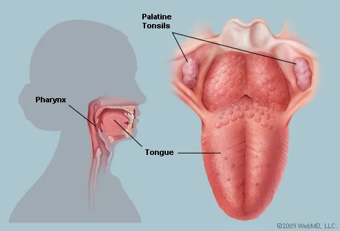
  • sore throat
  • fever
  • chills
  • muscle ache
  • fatigue
In more advanced stages of HIV, AIDS is usually diagnosed and includes:

  • White spots on the tongue or mouth
  • blurred vision
  • shortness of breath
  • diarrhea
  • swollen glands
Pawlina, Wojciech. "Connective Tissue." Histology A Text and Atlas By Michael H. Ross. 6th ed. N..: n.p., n.d 455 Print.               The two main contributors listed for the 6th edition of this textbook, Wojciech Pawlina and Michael H. Ross.
"What Is AIDS? What Is HIV?" Medical News Today. MediLexicon International, 13 May 2012. Web. 27 Oct. 2013.