Sunday, December 8, 2013

My Dictionary: Urinary System

Cortical labyrinths- the region between medullary rays contains renal corpuscles

http://www.dartmouth.edu/~anatomy/Histo/lab_5/renal/DMS152/popup.html

Bowman's capsule- double layered epithelial cup 

http://www.ouhsc.edu/histology/text%20sections/urinary.html
 Juxtamedullary nephrons- 1/8 of total nephron count. Contain long loops of Henle that go into the inner region of the pyramid.

http://faculty.southwest.tn.edu/rburkett/A%26P2%20urinary_system.htm

Sunday, December 1, 2013

Reflections: Digestive System III

     The liver is surrounded by fibrous connective tissue that is called Glison's capusle. Exterior to the capsule is a serous covering, the visceral peritoneum. However, the visceral peritoneum is not found where the liver meets with the diaphragm and other organs. The liver has grooves that divided into four lobes, two large lobes (left and right lobe) and two smaller lobes (quadrate and caudate lobes). The liver starts off as an endodemal evagination in the embryo. Then it forms the hepatic diverticulum, which proliferates and create hepatocytes that constitute the parenchyma. The liver is important in producing and secreting plasma proteins, regulating vitamins and nutrients, and maintains blood glucose levels. Moreover, it also plays a role in degrading drugs and toxic substances. The liver acts as an exocrine gland because it produces bile secretion and lastly, it also functions as an endocrine gland.
     The gallbladder is located at the visceral surface of the liver. Its main function is to store bile, it also removes approximately 90% of the water. The small intestine secretes hormones that cause contractions of the gallbladder that lead to bile discharge into the duodenum. The pancreas is divided into three sections: head, body, and tail. The gland is surrounded by a capsule that is made up of a layer of connective tissue. The pancreas as both an endocrine and exocrine gland. The endocrine component of the gland regulates blood glucose levels by synthesizing and secreting insulin and glucagon. The exocrine component of the gland functions as a serous gland and secretes digestive enzymes. 

Wednesday, November 20, 2013

Salivary Gland Tumors

     Salivary gland tumors typically occur in the major salivary glands. The three major salivary glands are the parotid gland, submandibular gland, and sublingual gland. The minor glands are found throughout the mouth, pharynx, larynx, and paranasal sinuses. Most of the salivary tumors are benign, meaning that they are not cancerous. There are usually in the parotid gland or in the palate if it is a minor salivary gland tumor.
    Most of the benign tumors are pleomorphic adenoma tumors. This tumor has epithelial tissue with ducts and epithelial cells mixed with areas that have similar characteristics of ground substance of connective tissue. The myoepithelial cells are what produce the tissues that resemble connective tissue. Patients that have these benign tumors claim to have no pain in the affected glands. The reason for this may be that the innervated nerves are numb and weak. What should a patient do with a benign salivary gland tumor? Surgery is a possible option for parotid gland tumors in which the entire gland will be removed.
Pawlina, Wojciech. "Connective Tissue." Histology A Text and Atlas By Michael H. Ross. 6th ed. N..: n.p., n.d 555 Print.               The two main contributors listed for the 6th edition of this textbook, Wojciech Pawlina and Michael H. Ross.
http://emedicine.medscape.com/article/194522-overview

My Dictionary

Palatine tonsils- located on both sides of the oropharynx opening
http://www.webmd.com/oral-health/picture-of-the-tonsils
Tubal tonsils- located behind the auditory tube opening on the later walls
Lingual tonsil- located at the tongue's base

http://www.knowyourbody.net/tubal-tonsil.html

Pharyngeal tonsils- at the top of the nasopharynx
http://hanatomy.com/anatomy-directory/lingual-palatine-and-pharyngeal-tonsils/

Thursday, November 14, 2013

Reflections: Integumentary System


     As we closed the chapter dealing with the integumentary system, I decided it would be good to do a brief review on what we learned. The integumentary system is composed of two main layers: the epidermis and the dermis, and there are two types of skin (thin and thick). Thin skin has hair and is more prevalent as opposed to thick skin which is only found in the palms of the hands and the soles of the feet.
     The epidermis is the layer of skin that is exposed and is visible. The dermis lies directly beneath the epidermis. Then there is the hypodermis that contains adipose tissue and connective tissue. The epidermis is further divided into 5 layers: stratum corneum, stratum lucidum, stratum granulosum, stratum spinosum, and stratum basale. The stratum corneum is the layer that varies in thick skin.
     The cells of the epidermis are keratinocytes, melanocytes, Langerhans', and Merkel's cells. The epidermis is made up of keratinized stratified squamos epithelium. The keratinocytes are the cells that function to separate the organism from the outside environment. Melanocytes are the cells that secrete melanin. Melanin is important because it is the reason that we are protected from UV radiation. It also produces the pigment of the epidermis. Langerhans' cells are the one's that present antigen's to the immune system. Lastly, Merkel's cells function in stratum basale of the epidermis and they are the most abundant in the fingertips and other places that sensory perception is intense.
http://classroom.sdmesa.edu/eschmid/Chapter5-Zoo145.htm

Thursday, October 31, 2013

Encounters: Urinary Tract Infection

      
     After finishing the lymphatic system chapter and turning in our disease projects this week, I felt motivated to write about a situation that happened to my friend. Earlier this week, she was experiencing back pain and had a burning sensation during urination. She was soon diagnosed with a urinary tract infection. Since she is my friend, I was worried about her and felt compelled to research the disease itself, the causes, and the symptoms. 
     In my research I discovered that UTI's are basically bacteria that has infected the urinary system. What entirely does the urinary system comprise? It consists of the kidney's, ureters, bladder, urethra, and sphincter muscles. Since women have short urethra's they are more susceptible to getting a UTI than men. Causes vary greatly and range from having sexual intercourse to pieces of feces coming into contact with the urethra. It can happen whenever bacteria gains access to the urethra in any way or form. 
Symptoms may include the following:

  • cloudy urine
  • painful/ burning sensation when urinating
  • frequent need to urinate
  • back pain
  • abdominal pain
  • nausea
     Fortunately, there are antibiotics available that will help with the discomfort. However, after taking the antibiotics she did mention that she felt extremely nauseous, dizziness, and wanted to vomit. She is now well and not feeling any discomfort. 


http://www.medicalnewstoday.com/articles/189953.php
http://www.mayoclinic.com/health/urinary-tract-infection/DS00286/DSECTION=symptoms
http://www.plannedparenthood.org/health-topics/womens-health/utis-4283.htm

Tuesday, October 29, 2013

Human Immunodeficiency Virus

     HIV is a retrovirus and it attacks the immune system by invading T helper cells. It contains an enzyme called reverse transcriptase that aids it in making DNA with the RNA that it has. After several years, approximately 11 years, clinical symptoms of AIDS will start to appear. This is because HIV is also a lentivirus, which basically means that it is a slow occurring virus. The HIV molecule has gp 120 proteins surrounding the molecule and bind to CD4 proteins on the T helper cell. This is rather strange because it seems as if the CD4 protein acts as a receptor for the HIV molecule. Once the HIV molecule is inside the T helper cell, it injects all of its infected material. The T helper cell ends up making copies of the HIV molecule and the immune system responds by producing cytotoxic CD8 T cells. This ultimately results in a war for the immune system because it fights itself between the virus infected CD4 T cells and the cytotoxic CD8 cells. There are various ways that HIV can be transmitted from one person to another.
For example:

  •  anal sex
  •  oral sex
  •  vaginal sex
  •  Exchange of blood 
  •  Breast feeding 
     The reason these are all ways in which HIV can be transmitted is because HIV is found in semen, vaginal fluids, blood, and breast milk. Therefore, it can also be transmitted in other uncommon situations. For example, there have been cases where healthcare workers had been dealing with blood that was HIV positive and acquired the disease. Healthcare workers must be take proper precautions because it is critical for their life and the patient's.

Signs and Symptoms
In early stages of HIV signs and symptoms may include flu- like symptoms such as

  • sore throat
  • fever
  • chills
  • muscle ache
  • fatigue
In more advanced stages of HIV, AIDS is usually diagnosed and includes:

  • White spots on the tongue or mouth
  • blurred vision
  • shortness of breath
  • diarrhea
  • swollen glands
Pawlina, Wojciech. "Connective Tissue." Histology A Text and Atlas By Michael H. Ross. 6th ed. N..: n.p., n.d 455 Print.               The two main contributors listed for the 6th edition of this textbook, Wojciech Pawlina and Michael H. Ross.
"What Is AIDS? What Is HIV?" Medical News Today. MediLexicon International, 13 May 2012. Web. 27 Oct. 2013.

Wednesday, October 9, 2013

My Dictionary


Nervous tissue is definitely one intriguing subject as it is controls functions that begin with the external environment and get interpreted by the brain. Although, it is an interesting subject, there are some terms that get a little confusing as they are similar but location can differentiate them. For example:

Oligodendrocytes are cells of the central nervous system that work to support axons and create a myelin sheath to insulate the axon. The myelin sheath wraps around multiple axons and insulates them to guarantee the quick conduction of nerve impulses.
Schwann Cells have the same function, however, they are located in peripheral nervous system and only myelinate one cell.

There are three distinct characteristics of connective tissue in the peripheral nervous system:
Endoneurium which is an individual nerve fiber and the connective tissue surrounding it.
Perineurium is a nerve fascicle and the the connective tissue surrounding it.
Epineurium which is the connective tissue that wraps around a nerve and also fills the spaces that lie between the nerve fascicles. 


http://confessionsofaleedsstudent.blogspot.com/2012/06/neuroanatomy.html
http://houseofmind.tumblr.com/post/729689108/ever-wonder-what-comprises-a-nerve-nerves-have
http://classroom.sdmesa.edu/eschmid/Chapter10-Zoo145.htm


Monday, October 7, 2013

Encounters: Runners and Muscle Tissue

Young couple run together on a sunset


   While studying in the library a few days ago, I decided to reward myself with short study break and took a magazine from the bookshelf that was behind me. As I was skimming through it, I found an interesting article called Whatever Works. It was about a man who seems to enjoy running because he was devastated when he found out he might not be able to run ever again. Unfortunately, he injured his Achilles tendon while doing a six mile run one day. He looked up all sorts of treatments that he could do so that he could be up and running soon. It's ironic how he mentioned that websites such as WebMD and mayoclinic.com were too scientific for him to understand. Instead, he searched for blog sites. 
     This article helped me appreciate the wonderful function of muscle tissue. Skeletal, smooth, and cardiac muscle tissue all work in a synchronized manner. The fibers of skeletal muscle contract causing muscle to shorten. The contraction cycle is a fast and continuous cycle that involves different structures including: actin, myosin, troponin, tropomyosin, and myosin. It is initiated when a nerve impulse arrives at a neuromuscluar junction, then acetylcholine is released and causes depolarization of the cell. Calcium channels open and allow calcium to flow into the cell, bind to troponin, and permit tropomyosin to shift so that myosin can bind to actin and create a cross- bridge. As nerve impulses continue to depolarize the cell, contraction will continue. 


http://www.nutricisedr.com/publications/nutritional-misfits-10-bad-habits-of-runners/
http://people.eku.edu/ritchisong/301notes3.htm

Monday, September 30, 2013

Dupuytren's Disease

Dupuytren's Disease 

Dupuytren's disease is also widely known as Dupuytren's contracture because in certain progressed cases, the person develops a contracture in the hand causing the fingers to bend. Dupuytren's disease occurs when tissue that is located beneath the palm thickens. As a result, in severe cases, the person is unable to fully extend their fingers. When the connective tissue continues to contract it can lead to hypertrophic scar formation. Dupuytren's disease is typically found in Northern European males that are above the ago of 50. The cause is not known, but is thought to be hereditary in some cases.


Kenny, Tim. "Dupuytren's Contracture." Patient.co.uk, n.d. Web. 30 Sept. 2013. <http://www.patient.co.uk/health/dupuytrens-contracture-leaflet>. 
The author is Dr. Tim Kenny, their contact information is not available
. It was published through patients.co.uk. The author writes a few opinions such as when he stated, "Dupuytren's contracture is not usually painful." When it was first posted is not listed but it is updated regularly, the last update was June 15, 2013. The website contains mainly text and one picture of a hand that helps viewers get a visual image of what the disease looks like in an affected hand.

Pawlina, Wojciech. "Connective Tissue." Histology A Text And Atlas. By Michael H. Ross. 6th ed. N.p.: n.p., n.d. 183. Print.
The two main contributors listed for the 6th edition of this textbook are, Wojciech Pawlina and Michael H. Ross.  

Friday, September 27, 2013

Blood Running Through Your Veins


Leukocytes
There are five different types of leukocytes:


         (granulocytes)
  •  Basophils
  • Neutrophils
  • Eosinophils
        (agranulocytes)
  • Lymphocytes
  • Monocytes.
They can be divided into two separate groups: granular and agranular. These granules can be found in the cytoplasm of the cell. However, both granulocytes and agranulocytes contain a small portion of azurophilic granules which can also be identified as lysosomes. Each of the five cells have unique distinguishing features that make them easier to identify amongst each other.
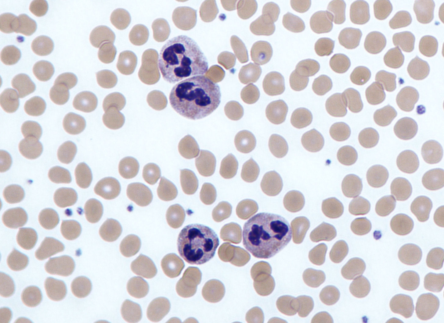
Neutrophils

These are the most common type of white blood cell's and granulocytes consisting of approximately 60-70% of leukocytes. Their multilobed nucleus makes it easy to distinguish them from the other leukocytes. This multilobed feature  gives them their other name, polymorphonuclear neutrophils. They are part of the granular cells and contain three types of granules: specific granules, azurophilic granules, and tertiary granules. The granules help determine their phagocytic function. They are motile cells which is important for them because they are the cells that first respond to tissue damage. Neutrophils contain receptors on their cell membrane, these receptors aid the cell in recognizing antigens. Once foreign substances are recognized phagocytosis can occur.
http://en.wikipedia.org/wiki/Neutrophil_granulocyte

Eosinophils

Eosinophils average the same size as neutrophils and consist of about 2-4% of leukocytes. They are granulocytes and contain two types of granules: specific and azurophilic granules. They have a bilobed nucleus with heterochromatic adjacent to their nuclear envelope and euchromatin in the nucleus. Eosinophils are created in the bone marrow then travel to connective tissue via peripheral blood. 

http://microanatomy.net/blood/more_eosinophils.htm

Basophils

Basophils are also similar in size to neutrophils. They are rarely found because they occupy roughly 0.5% of leukocytes. The lobed nucleus can be hard to find because of the numerous granules in the cell. Basophils also have two types of granules: specific and azurophilic granules. Their function is similar to those of mast cells. Basophils are created in bone marrow then released into peripheral blood where they migrate as a mast cell precursor. It then has the ability to become a mast cell. Basophils secrete mediators of inflammation such as, histamine, heparin, and heparin sulfate along with other mediators.
http://www.cytochemistry.net/microanatomy/blood/more_basophils.htm
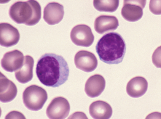
Lymphocytes

Most abundant type of agranulocytes and consist of 30% of leukocytes. There are three sizes of lymphocytes: small, medium, and large. The small cells are the most commonly found (90%). There are three functionally different cell types: T cells, B cells, and Natural Killer cells. T cells can be found in the thymus, they are so named T cells because they have T- cell receptors that function as cell- surface recognition proteins. B cells are active in producing antibodies. NK cells, as the name suggests, are involved in killing virus- infected cells and tumor cells. 

http://www.pathologystudent.com/?p=2186

Monocytes 

Monocytes are phagocytic cells and are the largest type of leukocyte. They maintain about 1-5% of white blood cells. They move from the bone marrow to the bloodstream where they stay for about 3 days before migrating to different sites of the mononuclear phagocytic system. The nucleus of the monocytes is slightly indented and contains centrioles and a developed Golgi apparatus. Monocytes are the second cells that appear at sites of inflammation where they develop into a macrophage. 


http://faculty.une.edu/com/abell/histo/histolab3a.htm

Having knowledge about these various kinds of leukocytes and their functions is beneficial. For example, neutrophils use receptors during phagocytosis such as toll -like receptors. Toll- like receptors release a fever- causing agent called pyrogen. Prostaglandins are synthesized and produce a fever by acting on the thermoregulatory center of the hypothalamus located in the brain. Therefore, a fever is the bodies natural defense to pathogens trying to invade.


Friday, September 6, 2013

My Dictionary

Connective Tissue 
- structurally and functionally underlies and supports epithelium.
There are three different types of CT: CT proper, specialized, CT, supporting CT, and embryonic CT

Connective Tissue Proper
http://histologyolm.stevegallik.org/node/99
 Loose connective tissue- has loosely arranged collagen fibers and numerous cells


http://medcell.med.yale.edu/histology/connective_tissue_lab/dense_regular_connective_tissue.php

Dense connective tissue- has arrange collagen fibers and few fibroblasts.
Dense CT is substantially stronger than loose CT and has two types: regular and irregular. 


Specialized Connective Tissue 
Specialized CT is categorized in three different types: adipose, lymphatics, and hematopoietic. 

Supporting Connective Tissue 
Supporting CT is characterized by any material that is in association with collagen, such as bone and cartilage. 

Bone tissue


http://www.nytimes.com/imagepages/2007/08/01/health/adam/1679BoneTissue.html


Embryonic Connective Tissue
Embryonic Tissue is made in the mesoderm layer. The two types are: mesenchyme and mucous CT

Mucous tissue

http://tae40905new.blogspot.com/2012_07_01_archive.html